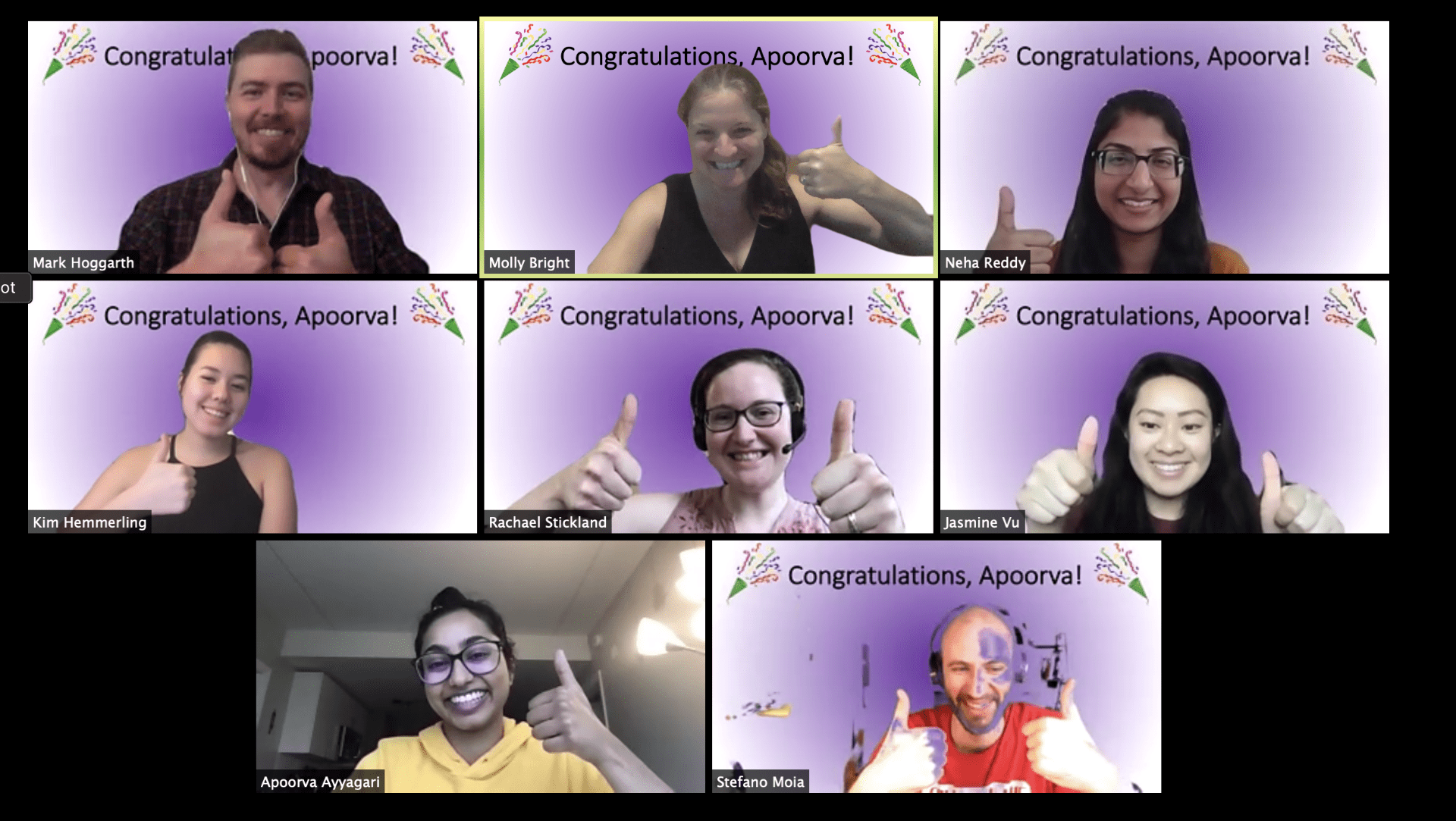
ANVIL welcomes two new Biomedical Engineering students to the lab!
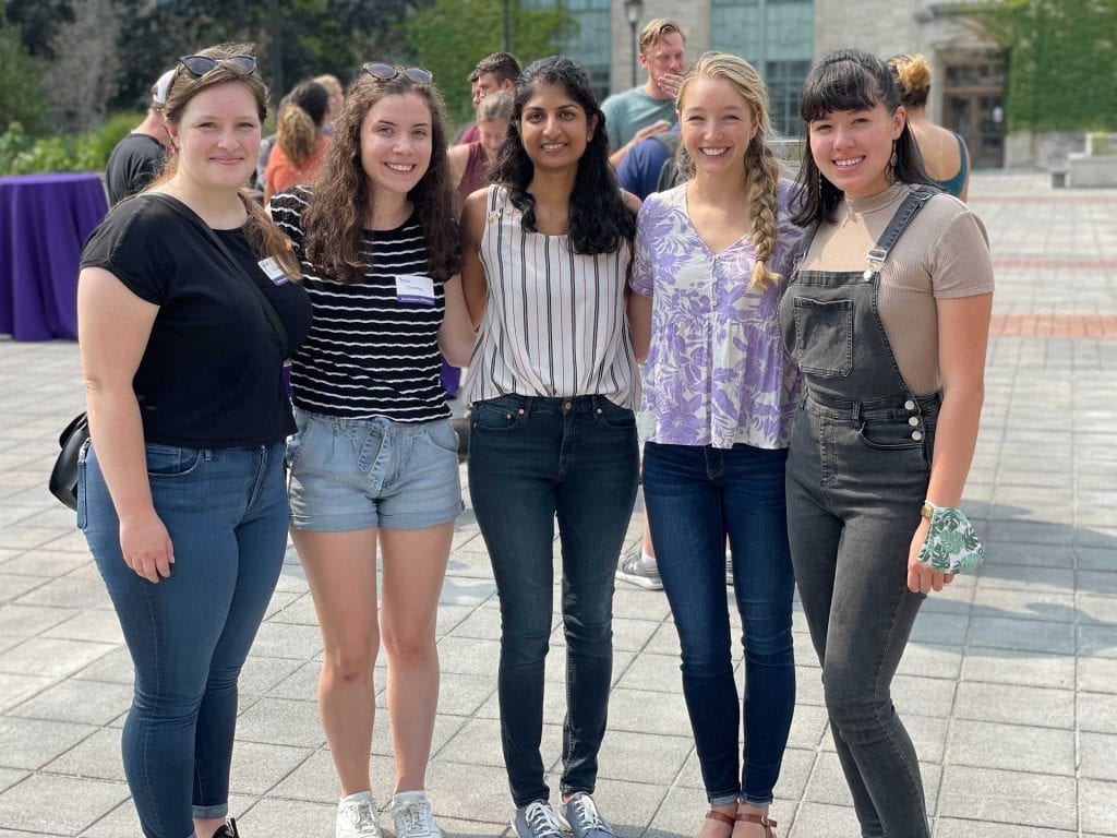
From left to right: Hannah and Becca join Neha, Kristina, and Kim as ANVIL BME doctoral students
Rebecca Clements is a new PhD student coming to us from the University of Delaware, where she completed her Bachelors degree in Biomedical Engineering. She has extensive research experience working in the Mechanical Neuroimaging lab at UD, where she used magnetic resonance elastography measures to better understand age-related changes to the brain.
Hannah Johnson, also an incoming PhD student, joins us from the University of Arizona, where she earned her Bachelors degree in Biomedical Engineering. As an undergraduate, she worked with Dr. Elizabeth Hutchinson in the Multi-Scale Brain Imaging Lab to identify longitudinal biomarkers of traumatic brain injury in preclinical models using MRI.
Becca and Hannah are excited to be joining the team, and we look forward to working with them!
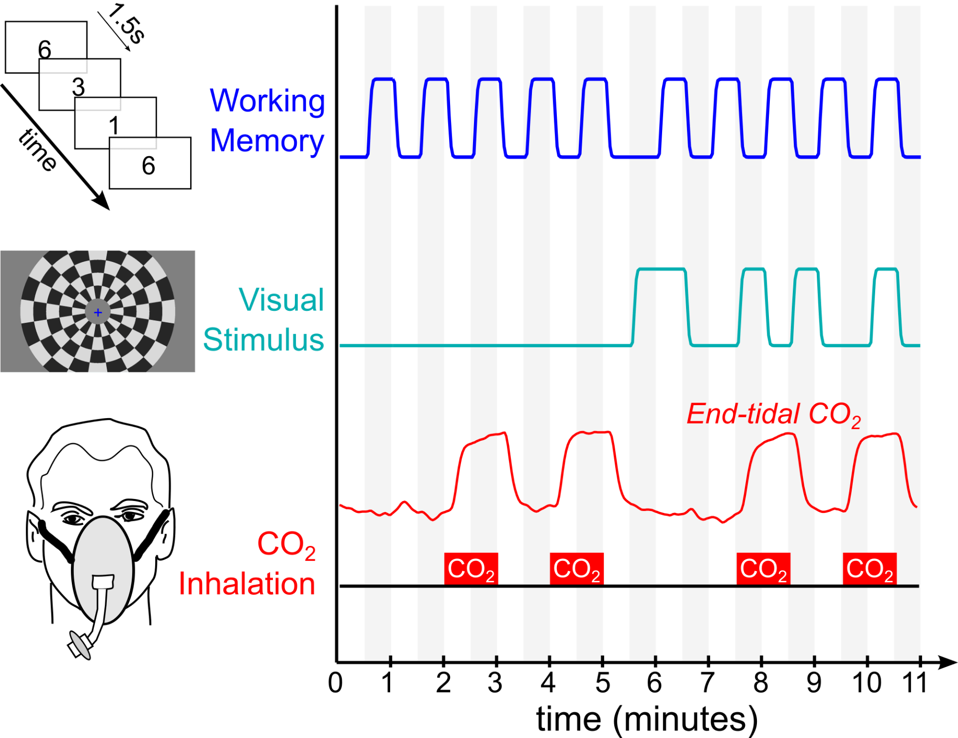
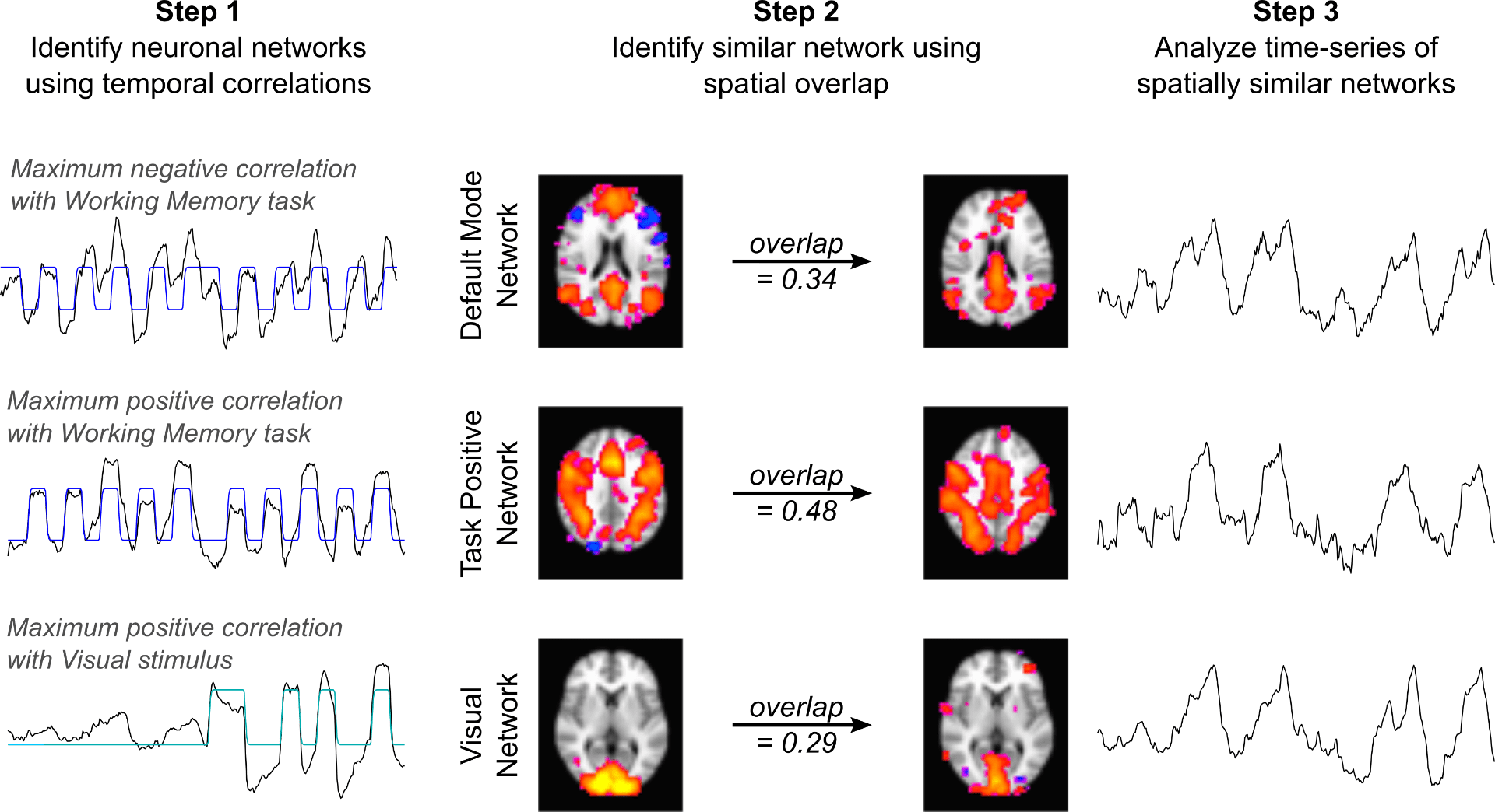
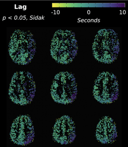 Congratulations to postdoctoral research fellow Rachael Stickland and colleagues on our publication in Neuroimage, titled


 The Craig H. Neilsen Foundation has awarded Dr. Molly Bright a SCIRTS Pilot Grant to use imaging to characterize neurovascular plasticity in spinal cord injury. Working with Dr. Milap Sandhu, PT PhD at the Shirley Ryan Ability Lab, we will employ fMRI and cerebrovascular imaging techniques to characterize how one session of Acute Intermittent Hypoxia impacts brain and spinal cord physiology. Acute Intermittent Hypoxia is an emerging intervention that transiently improves motor function in individuals with spinal cord injury; although large clinical trials are getting underway, we aim to better understand the mechanisms for these functional improvements in order to optimize or tailor the intervention. This study will span two years, and recruitment is soon beginning for individuals with incomplete cervical spinal cord injury as well as uninjured control participants. Mark Hoggarth, DPT (left) will be joining the lab later this fall to take the lead on spinal cord imaging and analyses. Welcome Mark, and stay tuned for details on how to get involved with the study!