 Graduate degrees were conferred for 5 students (now alumni!) of the Bright lab this morning. Drs. Kimberly Hemmerling, Neha Reddy, and Kristina Zvolanek were hooded and received their doctoral degrees, and Noah Northrop, MS, and Zac Zou, MS, received their master’s degrees after successful recent defenses! Congratulations to all the graduates!
Congratulations to Dr. Kimberly Hemmerling!
 Please join us in belatedly congratulating Dr. Kimberly Hemmerling on the successful defense of her PhD in Biomedical Engineering in April! Her disssertation, entitled Advanced functional MRI mapping of human spinal cord functional anatomy and vascular physiology, has made huge contributions to our functional understanding of the human spinal cord and the tools available to us to assess the spinal cord non-invasively. Her work surmounts the numerous challenges facing spinal cord imaging to map spinal cord vascular reactivity and motor activity, enabling understanding of spinal cord function at an individual level and advancing its utility for the functional assessment of clinical populations.
Kim has been an incredible asset to the Bright lab throughout her tenure, and her work has shaped the research directions that the lab is pursuing and will continue to influence ongoing and future projects. She’s served as an extraordinary leader in the lab, closely mentoring numerous undergraduate students and younger PhD students as they dive into research. Kim’s expertise and commitment to fostering a supportive and productive lab community will be greatly missed as she moves on to the next phase of her career, but wish her the best of luck with her next steps!
Congratulations to Dr. Neha Reddy!

We are thrilled to announce that Dr. Neha Reddy has successfully defended her doctoral dissertation, entitled Systems-level sensorimotor mapping with fMRI for applications in brain disorders! Neha’s research has significantly advanced sensorimotor mapping through innovative fMRI and signal-processing methodologies. Her work with multi-echo independent component analysis and whole-brain fMRI has improved understanding of somatosensory and motor neural systems and will advance knowledge of how these systems are impacted in neurological impairment such as stroke.
Throughout her time in the Bright Lab, Neha has demonstrated exceptional research skills, technical innovation, and leadership. Her work ethic and intellectual curiosity have been instrumental to our lab’s success, and have set a high standard that we will endeavor to live up to. We wish her best of luck as she returns to medical school this summer, and look forward to witnessing her continued contributions to the field of medicine as a physician-scientist! Please join us in congratulating Dr. Reddy on this remarkable achievement!
Congratulations to Dr. Kristina Zvolanek!
 Many congratulations to Kristina Zvolanek, who successfully defended her PhD in Biomedical Engineering on January 9! Her thesis, entitled Novel functional MRI approaches for mapping cerebrovascular hemodynamics in sickle cell disease, addresses critical gaps in understanding cerebrovascular complications in pediatric sickle cell disease. Her work has developed innovative functional MRI approaches to measure cerebrovascular reactivity and circulation time, creating new pathways for investigating hemodynamics in this vulnerable population.
As a founding member of the Bright Lab, Kristina has been instrumental in shaping lab culture, demonstrating incredible leadership, exceptional mentorship, and scientific rigor. Her contributions will continue to shape our ongoing projects and future work. We are incredibly proud of her and will miss her greatly, but we wish her the best of luck in her new role as a consultant with Beghou Consulting. Congratulations, Dr. Zvolanek!
New paper on mapping hand-grasp motor task activity in the spinal cord
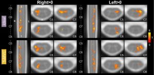 Hand-grasping is an important and clinically significant daily function, but more work is needed to understand the associated spinal cord neural activity. In this study, participants performed a hand-grasping task with both hands and spinal cord fMRI motor activation was modeled with two methods: (1) a typical idealized block design (Ideal) and (2) based on recorded grasp force normalized to individual maximum voluntary contraction (%MVC). Robust detection of hand-grasping activity was shown in the spinal cord, highly lateralized ipsilateral to the side of the task. In addition, the impact of sample size, number of fMRI runs, and spatial smoothing on activation estimates was explored. Overall, we emphasize the importance of a task that is well-controlled within and across participants. Using individually calibrated tasks in people with motor impairments will be especially beneficial to compare across groups.If you want to learn more, please check out the paper here!
Multiple ANVIL successes to celebrate!
Congratulations are in order for three outstanding accomplishments from ANVIL lab members this past month!
Becca Clements (2nd year PhD student in BME) was awarded a 3-year fellowship from the NSF Graduate Research Fellowship Program! This will fund her exploration into the organization of vascular regulation in the human brain, and particularly the presence of long-distance “vascular networks” that may support typical functional neural networks.
Kim Hemmerling (4th year PhD student in BME) received a stellar 6%ile score on her NIH F31 Predoctoral Fellowship application! This score reflects the amazing potential for the spinal cord fMRI methods she has been developing in our lab, and specifically towards looking at atypical upper limb movement patterns post-stroke.
And finally, Max Wang (Postbac Research Technologist) has been awarded a prestigious Fulbright Scholarship! Max will pursue his Masters research in translational neuroimaging at the University of Nottingham in the UK starting this fall.
These successes reflect the impressive ongoing hard work and perseverance of our trainee researchers. We are so pleased to see these efforts acknowledged and rewarded, and we are looking forward to seeing what new science emerges as they begin these exciting projects. Congratulations Becca, Kim, and Max!
Exploring variability in spinal cord fMRI processing
 How reliable are our current techniques for processing spinal cord fMRI data? In a recently published paper, Dr. Mark Hoggarth and Max Wang explore how person-to-person variability in the contouring of fMRI spinal cord masks affects downstream analyses. Using masks drawn by 8 raters of varying experience to inform co-registration to standard template space, they identify spatial differences in the accuracy of registration related to underlying image contrast and rater experience. At the group-level, spatial differences were present in activation maps although no systematic effect on overall activation level was identified. This work characterizes how variability in manually contoured masks propagates to results in spinal cord fMRI, demonstrating that standardization of processing pipelines and improving image acquisition should be prioritized.
This work was performed in collaboration with Dr. Kenneth Weber at the Stanford Systems Neuroscience and Pain Lab.
You can find the full access paper here!
New paper on the correlation between baseline blood flow and vascular reactivity in the brain
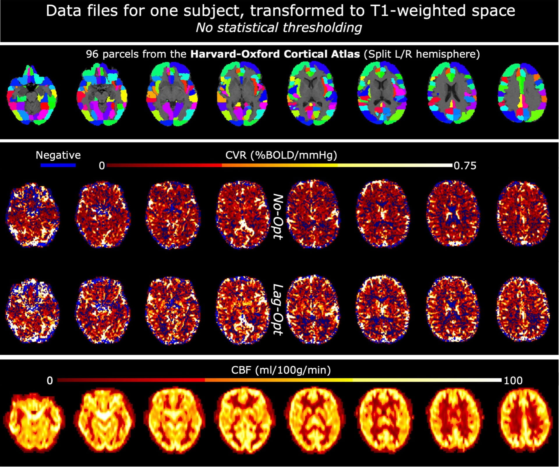 A recent paper from the lab, led by Dr. Rachael Stickland, identifies a positive correlation between baseline cerebral blood flow (CBF) and Blood Oxygenation Level Dependent cerebrovascular reactivity (BOLD-CVR), measured with MRI. This positive correlation is seen between and within subjects and is strengthened by the addition of a breathing task to a resting-state acquisition and by optimizing for hemodynamic lag effects in CVR modeling. This work demonstrates how two analytical factors affect the observed relationship
 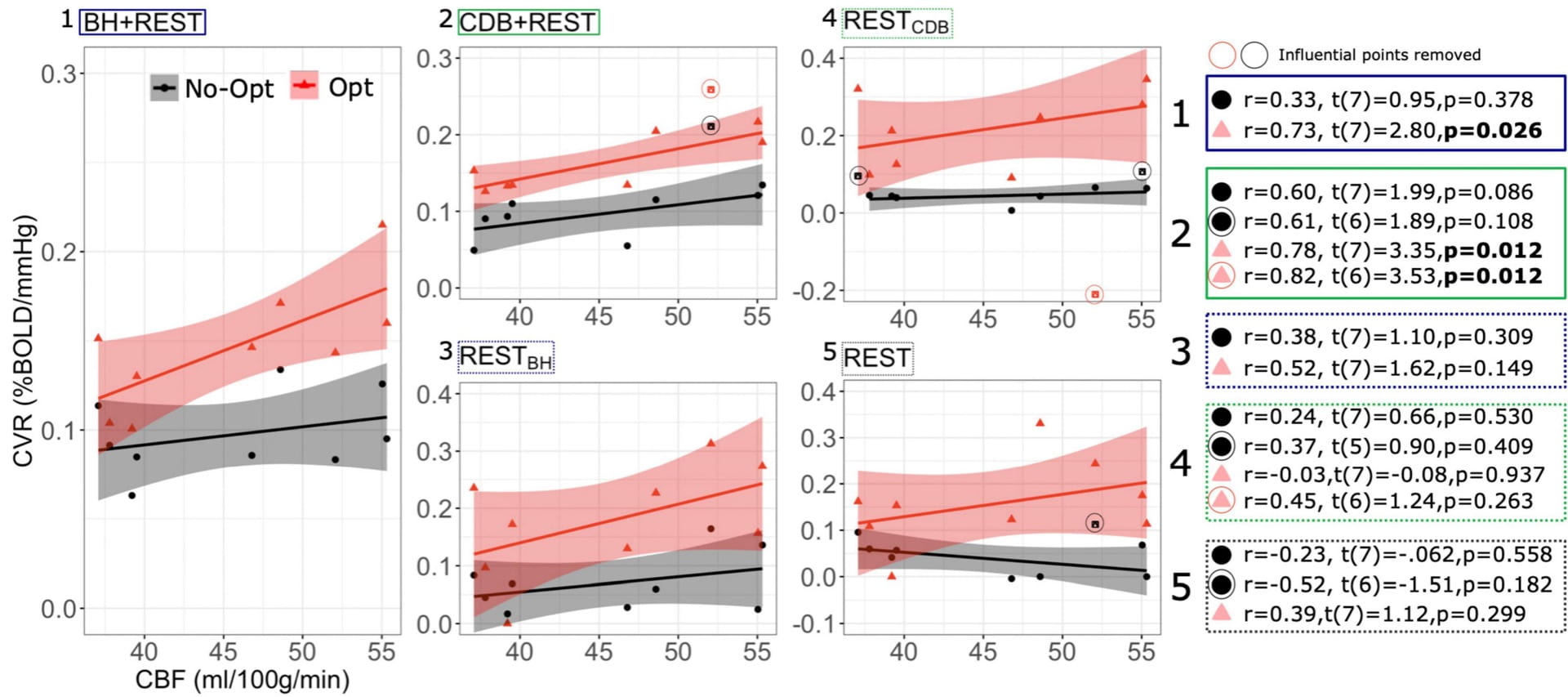 between baseline CBF and BOLD-CVR in healthy populations; the relationship becomes even more relevant to understand when interpreting results in
populations with altered vascular baselines or impaired cerebrovascular reserve.
This work was performed in collaboration with Dr. César Caballero-Gaudes’ lab at the Basque Center on Cognition, Brain and Language in Spain.
Check out the paper here!
Conference paper on visualization tool for assessment of spinal cord fMRI data quality
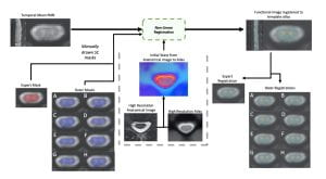
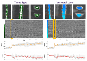 Check out our conference paper that was presented at the IEEE Engineering in Medicine and Biology Conference and published online in December 2021! This paper details our method, developed by PhD candidate Kimberly Hemmerling, for visualization of complex spinal cord fMRI data as heatmaps. This work was inspired by a visualization technique designed for brain fMRI data (Power et al., 2017). Our heatmaps can be visualized alongside motion or physiological traces that may be used to identify coincidence of signal variation. The technique may be easily integrated into a preprocessing pipeline to visualize the effect of a preprocessing step, identify spatially or temporally varying artifacts, or to assess general data quality.Check out the work here, and our GitHub repository (BrightLab-ANVIL/spinalcordplot), which contains the code and example data necessary to generate the plots.
Lab awarded NIH funding for clinical trial to enhance brain blood flow
Molly has been awarded a two-year R21 grant from NINDS to determine the ability of Acute Intermittent Hypoxia therapy to enhance blood flow in the brain of healthy adults. This controlled cross-over trial is an exploratory “high-risk, high-reward” study that examines whether a simple intervention can increase resting cerebral perfusion and the vasodilatory responsiveness of cerebral blood vessels. The intervention is already used in several studies at Northwestern University and the Shirley Ryan AbilityLab to evoke neural plasticity for rehabilitation, and this study explores whether it also achieves vascular plasticity. Participants will undergo three weeks of regular, brief exposures to hypoxia, and advanced arterial spin labeling MRI techniques and MRI-compatible gas challenges will be used to assess the impact on the cerebrovasculature. Recruitment starting soon; see the Current Studies page for more information!