Current Research Studies Recruiting Participants
We have numerous ongoing studies, in both healthy control populations and specific clinical groups, and we are always looking for participants interested in getting involved! For details and contact information, check out this page with instructions on how to get in touch for more information.
Results from Lab Research Studies
Research from our lab leads to methodological developments and new understanding of human physiology and function – we’ve started a new page with short summaries of key research findings, which we will update as current studies bear fruit.
Resources
Advanced Methods for Cleaning up fMRI Time-Series
Educational course from the Organization for Human Brain Mapping annual meeting, 2017, Vancouver.
Organizers: Molly Bright and Kevin Murphy; Faculty: Cesar Caballero Gaudes, Daniel Handwerker, Jonathan Power, Molly Bright, Ludovica Griffanti, Prantik Kundu

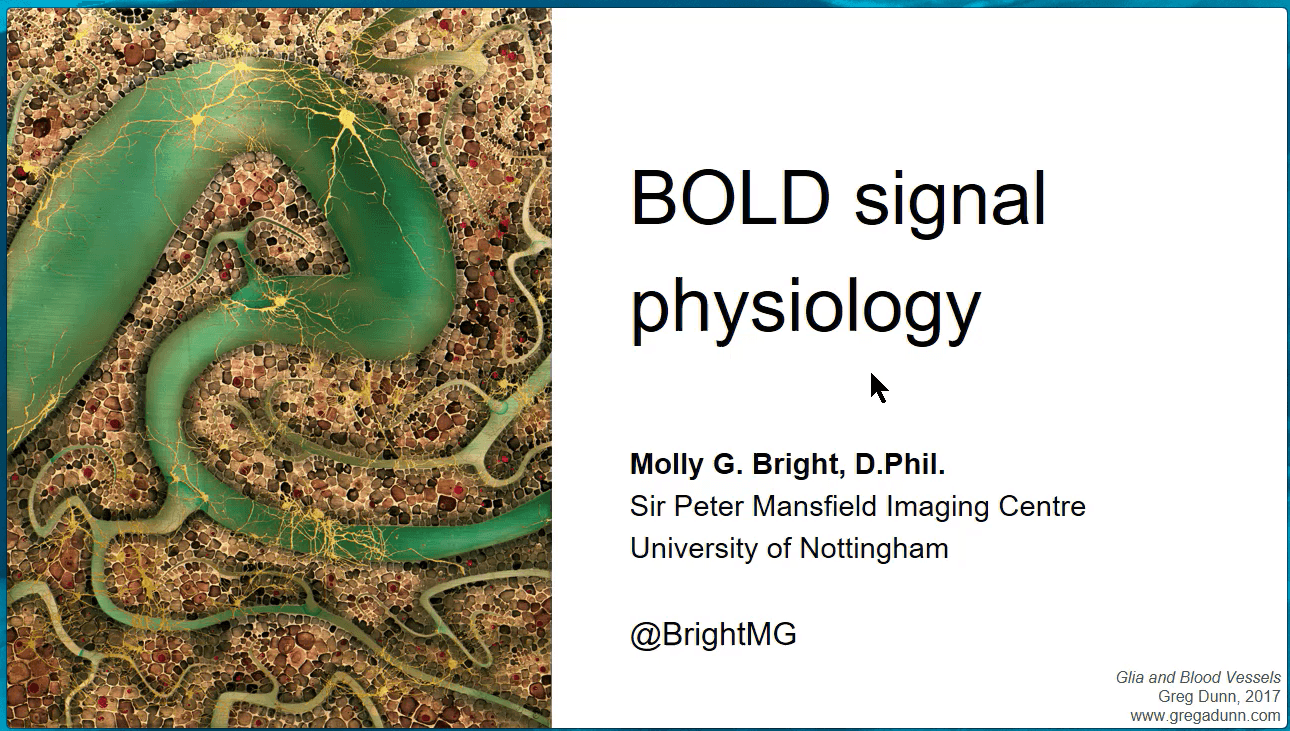
BOLD Signal Physiology
Educational presentation at the ISMRM annual meeting, 2017, Honolulu.
Seven Minutes of Science Research Presentations
Research presentations given by several of our past and current graduate students as part of Northwestern’s Research Communication Training Program.
Publications
Published Articles
Hemmerling KJ, Vigotsky AD, Glanville C, Barry RL, Bright MG. (2025) Data-driven denoising in spinal cord fMRI with principal component analysis. bioRxiv [Preprint].
Banerjee R, Kaptan M, … Hemmerling KJ, Mobarak-Abadi, Hoggarth MA, Howard MA, Bright MG et al. (2025) EPISeg: Automated segmentation of the spinal cord on echo planar images using open-access multi-center data. bioRxiv [Preprint].
Clements RG, Zvolanek KM, Reddy NA, Hemmerling KJ, Bayrak RG, Chang C, Bright MG. (2024) Quantitative mapping of cerebrovascular reactivity amplitude and delay with breath-hold BOLD fMRI when end-tidal CO2 quality is low. bioRxiv [Preprint].
Johnson HR, Wang MC, Stickland RC, Chen Y, Parrish TB, Sorond FA, Bright MG. (2024) Variable Cerebral Blood Flow Responsiveness to Acute Hypoxic Hypoxia. bioRxiv [Preprint].
Zvolanek KM, Moore JE, Jarvis K, Moum SJ, Bright MG.(2024) Macrovascular blood flow and microvascular cerebrovascular reactivity are regionally coupled in adolescence. Journal of Cerebral Blood Flow & Metabolism.
Hemmerling KJ, Hoggarth MA, Sandhu MS, Parrish TB, Bright MG. (2024) MRI mapping of hemodynamics in the human spinal cord. bioRxiv [Preprint].
Comparing end-tidal CO2, respiration volume per time (RVT), and average gray matter signal for mapping cerebrovascular reactivity amplitude and delay with breath-hold task BOLD fMRI. Neuroimage. 272:120038.
Effects of variability in manually contoured spinal cord masks on fMRI co-registration and interpretation. Front Neurol. 13:907581.
Lag-Optimized Blood Oxygenation Level Dependent Cerebrovascular Reactivity Estimates Derived From Breathing Task Data Have a Stronger Relationship With Baseline Cerebral Blood Flow. Front Neurosci. 16:910025.
Hemmerling K, Bright MG (2021) A visualization tool for assessment of spinal cord functional magnetic resonance imaging data quality. Accepted in Proceedings of the 43rd Ann. Int. Conf. of IEEE Eng Med Biol Soc (EMBC), virtual meeting.
Levitis E, Gould van Praag, CD, Gau R, Heunis S, DuPre E, Kiar G, Bottenhorn KL, … Bright MG et al. (2021) Centering inclusivity in the design of online conferences-An OHBM-Open Science perspective. GigaScience 10(8):giab051.
Stickland RC, Zvolanek KM, Moia SC, Ayyagari A, Caballero-Gaudes C, Bright MG (2021) A practical modification to a resting state fMRI protocol for improved characterization of cerebrovascular function. Neuroimage 239:118306.
Vu J, Nguyen BT, Bhusal B, Baraboo J, Rosenow J, Bagci U, Bright MG, Golestanirad L. (2021) Machine learning-based prediction of MRI-induced power absorption in the tissue in patients with simplified deep brain stimulation lead models. IEEE Transactions on Electromagnetic Compatibility 63(5):1757–1766.
Moia S, Termenon M, Uruñuela E, Chan G, Stickland RC, Bright MG, Caballero-Gaudes C (2021) ICA-based denoising strategies in breath-hold induced cerebrovascular reactivity mapping with multi echo BOLD fMRI. Neuroimage 223:117914.
Editorial: Imaging Cerebrovascular Reactivity: Physiology, Physics and Therapy. Front Physiol. 12:740792.
Pinto J, Bright MG, Bulte DP, Figueiredo P (2021) Cerebrovascular Reactivity Mapping Without Gas Challenges: A Methodological Guide. Front. Physiol. 11:608475.
Bright MG, Whittaker JR, Driver ID, Murphy K (2020) Vascular physiology drives functional brain networks. Neuroimage 217:116907.
Moia S, Stickland RC, Ayyagari A, Termenon M, Caballero-Gaudes C, Bright MG (2020) Voxelwise optimization of hemodynamic lags to improve regional CVR estimates in breath-hold fMRI. 42nd Annual International Conference of the IEEE Engineering in Medicine & Biology Society (EMBC), Montreal, QC, Canada, 2020, pp. 1489-1492.
Whittaker JR, Driver ID, Venzi M, Bright MG, Murphy K (2019) Cerebral autoregulation evidenced by synchronized low frequency oscillations in blood pressure and resting-state fMRI. Frontiers in Neuroscience 13:433.
Dury RJ, Falah Y, Gowland PA, Evangelou N, Bright MG, Francis ST (2019) Ultra-high-field Arterial Spin Labeling MRI for non-contrast assessment of cortical lesion perfusion in Multiple Sclerosis. Eur. Radiol. 29(4):2027-2033.
Bright MG, Croal PL, Blockley NP, Bulte DP (2019) Multiparametric measurement of cerebral physiology using calibrated fMRI. Neuroimage 187:128-144.
Whittaker JR, Bright MG, Driver ID, Babic A, Murphy K (2019) Changes in cerebral arterial blood volume during lower body negative pressure measured with MRI. Neuroimage 187:166-175.
Bright MG, Tench CR, Murphy K (2017) Potential pitfalls in denoising resting state fMRI data using General Linear Models. Neuroimage 154:159-168.
Driver ID, Whittaker JR, Bright MG, Muthukumaraswamy, S, Murphy K (2016) Arterial CO2 fluctuations modulate neuronal rhythmicity; implications for MEG and fMRI studies of resting state. J Neurosci 36(33):8541-50.
Tewarie P, Bright MG, Hillebrand A, Robson SE, Gascoyne LE, Morris PG, Meier J, Van Mieghem P, Brookes MJ (2016) Predicting haemodynamic networks using electrophysiology: the role of non-linear and cross-frequency interactions. NeuroImage 230:273-292.
Bright MG, Murphy K (2015) Is fMRI “noise” really noise? Resting state nuisance regressors remove variance with network structure. Neuroimage 114:158-169.
Whittaker J, Driver I, Bright MG, Murphy K (2015) The absolute CBF response to activation is preserved during elevated perfusion: implications for neurovascular coupling measures. Neuroimage 125:198-207.
Bright MG, Bianciardi M, de Zwart JA, Murphy K, Duyn JH (2014) Early anti-correlated BOLD signal changes of physiologic origin. Neuroimage 87:287-296.
Bright MG, Murphy K (2013) Reliable quantification of BOLD fMRI cerebrovascular reactivity despite poor breath-hold performance. Neuroimage 83:559-568.
Bright MG, Murphy K (2013) Removing motion and physiological artifacts from intrinsic BOLD fluctuations using short echo data. Neuroimage 64(1):526-537.
Bulte DP, Kelly M, Germuska M, Xie J, Chappell MA, Okell TW, Bright MG, Jezzard P (2012) Quantitative measurement of cerebral physiology using respiratory-calibrated MRI. Neuroimage 60:582–591.
Bright MG, Donahue MJ, Duyn JH, Jezzard P, Bulte DP (2011) The effect of basal vasodilation on hypercapnic and hypocapnic reactivity measured using magnetic resonance imaging. J Cereb Blood Flow Metab 31(2):426-38.
Bright MG, Bulte DP, Jezzard P, Duyn J (2009) Characterization of regional heterogeneity in cerebrovascular reactivity dynamics using novel hypocapnia task and BOLD fMRI. Neuroimage 48(1):166-75.
Editorials, Comments, and Book Chapters
Bright MG, Murphy K (2017) Cleaning up the fMRI time series: mitigating noise with advanced acquisition and correction strategies. Neuroimage 154:1-3.
Bulte DP, Bright MG, Howe F, Corfield D (2016) Vascular Response to Hypoxia and Hypercapnia. In: Bammer R ed. MR & CT Perfusion Imaging: Clinical Applications and Theoretical Principles. Wolters Kluwer. ISBN/ISSN: 9781451147155.