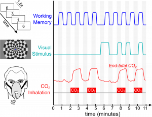
 Our most recent work demonstrating that the brain’s vasculature is regulated to mimic neural networks is now published in Neuroimage: https://doi.org/10.1016/j.neuroimage.2020.116907. This work was performed in collaboration with Prof. Kevin Murphy’s lab at Cardiff University Brain Research Imaging Center in the UK.
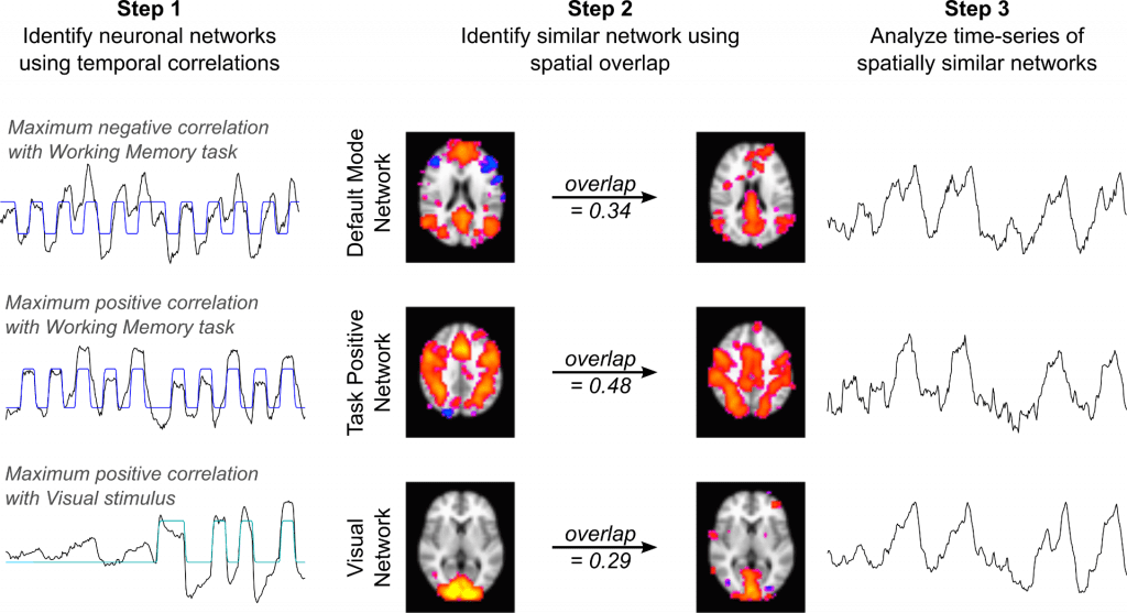
Using fMRI, we simultaneously administer two neural paradigms (a working memory task and a visual stimulus) and one “vascular” paradigm that dilates blood vessels systemically (inhaled CO2). We then averaged together 30 fMRI datasets and used Independent Component Analysis to decompose the average dataset into network components. We readily identify three “neural networks” that show strong temporal correlation with the neural stimulus paradigms. These represent the Default Mode Network, Task Positive Network, and Visual Network – three robust and commonly observed functional brain networks expected to be activated or deactivated by our neural paradigms. However, we also see three additional components with similar network structure, and these three networks predominantly reflect the vascular stimulus design.
Our results demonstrate, for the first time, pairs of spatially similar neural and vascular brain networks. This suggests that the brain’s vasculature may be regulated to support specific brain networks, which must be taken into account to interpret fMRI studies of functional connectivity.
